• +91 9544652090
  • support@cloudexrad.com
  • Working Hours - Mon - Sun: 12.00 am - 11.59 pm
What are you looking for?

Tag Archives: CT scan

ctchest teleradiology

Teleradiology Services Are Forerunners Among the Healthcare Providers to Adapt to Hybrid Work in Contemporary Times

The healthcare management faced a tough situation during a pandemic time and emerged victorious in extending the best care to COVID & Non-COVD patients as well as safeguarding its medical care providers (clinical & non-clinical); and with the understanding that technology-driven practices and contact-less patient care are the new norms and necessity. 

Today, medical technology integrated with deep learning and artificial intelligence is one of the major contributing factors. On-line consultation through telemedicine, teleradiology, drone services, e-pharmacy, remote working, and at home services are some of the examples contributing to reduced or no exposure. Radiology was among the forerunners that made a swift adaption to new norms and supported the system with remote working. The technology companies too supported the medical system’s functionality either in maintaining or increasing the efficiency of the operational team during the patient flow surge. 

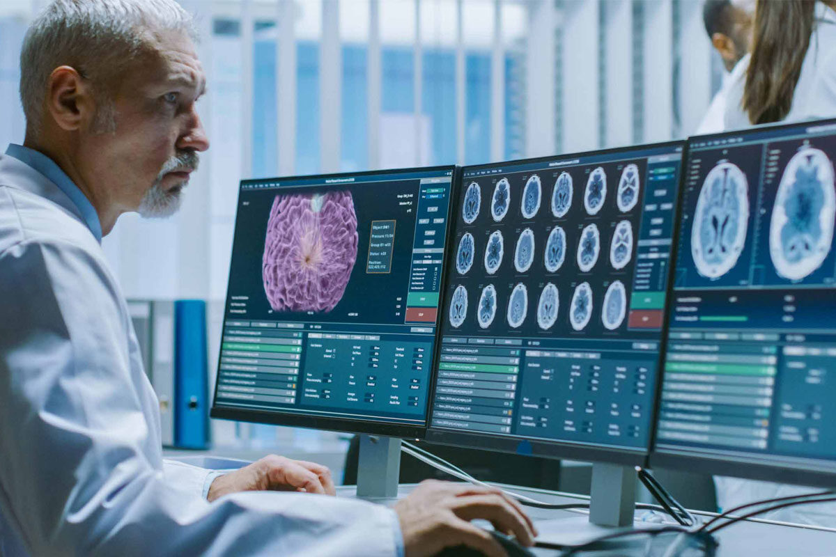
Teleradiology system driven by technological advancement has emerged as one of the most reliable modes of the patient care system in any given situation to deal with, be it a reporting for emergency or isolated COVID patients or IP/OPD patients, and could cope with the surge in image reporting through the option of remote reporting. This hybrid work norm supported many institutions in delivering patient care without any delay. Today’s teleradiology is integrated with the latest PACS, AI enabled workflow, internet connectivity with wide bandwidth and an experienced back-end support team that has made the reporting possible with short TAT. Technology companies have developed systems to transfer and archive the large dataset with integrity fulfilling the radiologist’s need for high-quality images for reporting. 

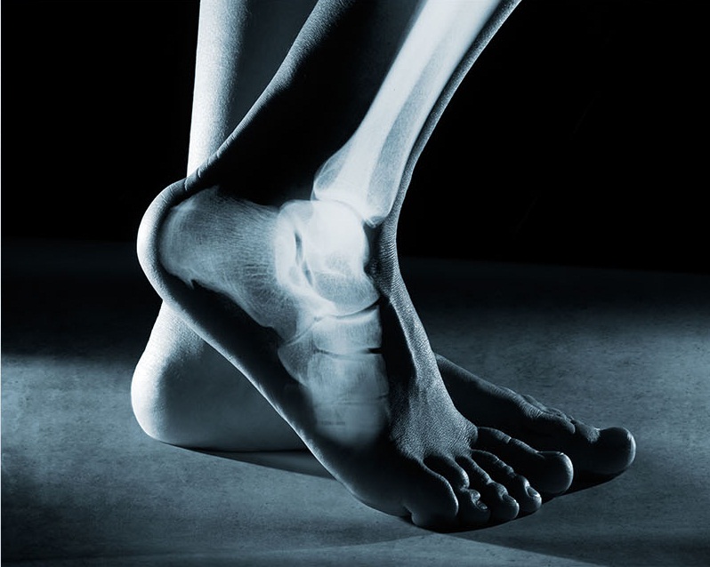
Machine Manufacturers are also contributing by developing systems that capture high-quality images with low dose radiation and high sensitivity. And to catch up with the trend of imaging services at point-of-care, compact mobile machines are getting developed to fulfill the need. The ultrasound machine and mobile X-ray machines are proving it with their wire-less and cloud-computing features. 

Remote Learning is being embraced by educators and many clinicians. The online recorded or live sessions have become the mode of learning for aspiring future physicians and medical professionals. Webinars/conferences and interactive sessions are being conducted virtually benefiting the healthcare providers and patients. 

With progressing time, the positive and productive approaches of radiologists, healthcare providers and technology companies the recovery from the pandemic is becoming a reality.  

Cloudex Teleradiology Solutions during Pandemic

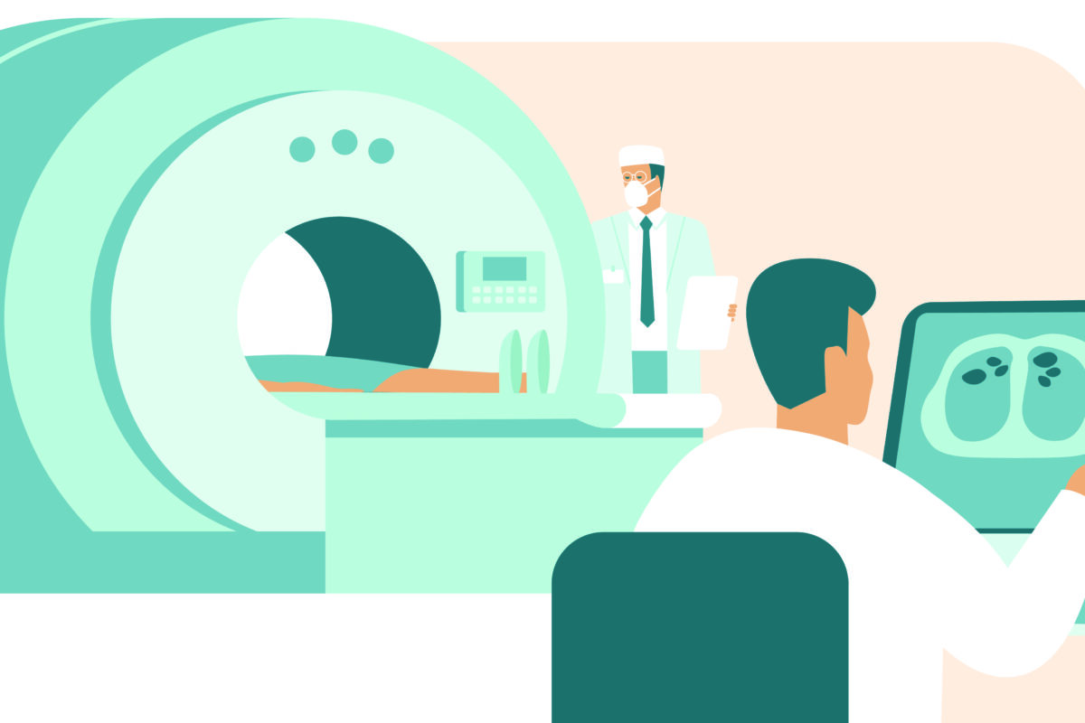
Cloudex with its advanced teleradiology service in India supported many health centers worldwide managing the patient surge with its prompt reporting services. The reporting was dominated by X-rays, CT and MRI scan mainly for emergency cases as other patients were curtailed from outpatient services. Our experienced and expert teleradiologists team extended their much needed services relentlessly round the clock. Safety measure guidelines were strictly followed by the back-end team while reporting was done remotely. The non-stop and timely service deliverance which includes subspeciality reporting in this crucial time helped many centers in facilitating patient recovery and saving lives and it turned out as their winning edge. 

At Cloudex, we support you to have that winning edge.

ct scan_workshop

5 Influencing Factors in Teleradiology Reporting

Radiology reporting with shorter TAT aided with tele reporting has brought about significant improvement in patient care outcomes in many departments of medical centers. TeleRadiology report plays a crucial role in terms of diagnosis and treatment planning. And sometimes, a few minutes of waiting period feels like long years and one wonders how to overcome those anxious moments of dilemma. Silence prevails on the patient’s side whereas an array of actions happens on the other side for radiology reporting. So, behind the complexity of radiological image interpretation lies an array of actions involving technology, protocols, coordination, communication, expertise which keep happening seamlessly. Many of these are visible in action status at the reporting stations or work stations of radiologists (on-site/off-site).

The radiologist/teleradiologist’s work-station is made up of hardware, software, and appropriate furniture & interiors. Factors like illumination in working space, ergonomics of chair & table to smallest of an object like a mouse which can possibly trigger the discomfort level to radiologists matters in the process. Briefing out these dynamic factors for understanding behind the scene actions in teleradiology –

1. Hardware:

  • The dual QHD monitors of size 21 / 30” diagonally equivalent to a 3 or 5 megapixel are used to view the images and write reports simultaneously. This monitor size reduces the need for repeated zooming in or panning out.
  • CPU with 8th generation, 16 GB RAM, 1GB GPU memory to match the speed of the image interpretation process of the radiologist.
  • V shaped keyboard and optic mouse with a scroll which contours the fit into adult hand providing the ergonomic posture for convenient working.
  • Bluetooth enabled headsets helps in audio and dictation.

2. Software:

  • PACS provides seamless transmission of images in correct order along with other required supporting health documents.
  • DICOM calibration to meet the DICOM standards which ensures the image displayed is perceptually linearized. This software also provides alerts for out-of-compliance issues through emails/notification.
  • Notification system alerting the radiologists on an upcoming critical case that needs urgent attention, TAT alarm, call out from other physicians for case discussion/clarification.

Communication platforms contribute in an immeasurable manner. The interaction between the reporting teleradiologist and referring physician is crucial and is a deciding factor and lifesaving in many cases. The alerts/notification / textual or audio messages etc have added speed to the reporting system.

  • TAT enhancing voice recognition services add lot of value to teleradiology services. It enables teleradiologist in engaging more time in image interpretation and sparing them from initial drafting of reports with features like Real-time self-editing, formatting, auto-punctuation, and electronic signature. It works as workflow accelerator helping in completing the greater number of image reporting in given time frame.

3. Furniture & Interiors:

  • The versatility of working space supports the radiologist’s long working hours. The ergonomics of the furniture like height of a table, lumbar supporting chair with adjustable seat height are important for comfortable viewing and reporting. This subtlety adds in promoting alertness and efficiency.
  • Luminance is given special care so as to safeguard radiologists from ocular distress and physical strain. The minimum standards of 350 ccd/m2 is maintained.

4. Connectivity & Server: Speed is the vital part because a lot of activity depends upon it – transmission, downloading, and storage-related rendering or archival of images, interpretation, and reporting of images.

  • A dedicated reliable high-speed internet connectivity is maintained. The minimum required internet speed is 512 KBPS-1MBPS whereas the speed of 4-8 MBPS is considered ideal enabling a smooth process for interpretation & reporting, and desirable speed is above 25 MBPS for interpretation of large image data set.
  • The whole process is supported through cloud computing or a quality-controlled web server

5. Cybersecurity: The infrastructure of PACS & DICOM are build keeping in mind the various guidelines provided by HIPPA/FDAs which are further integrated or upgraded with mitigation measures decided by the Teleradiology companies as company policy or SOP decided upon with their clients. Some of the well-known measures are selective encryption of the DICOM header/files, transport security, image de-identification, water-marking or digital signatures, and access to the system thru assigned user ID/password, etc.

Cloudex’s user-friendly work stations delivering customer satisfying results

At Cloudex, reporting stations are configured involving lots of detailing to ensure that teleradiologists are able to concentrate on interpretation and reporting without any distractions/interruptions and operational issues. The reporting platforms are integrated with various software like voice-recognition dictation, multimedia communication, notification apart from the latest PACS & case managing RIS. Importance is also given to illuminance, work-space comfort, sitting ergonomics, monitor configuration for maintaining teleradiologit’s optimal concentration and efficiency. We invest beyond infrastructure too for creating a conducive environment that is free from administrative burdens or logistical hurdles for teleradiologists. The organization’s emphasis on accuracy of reporting without a miss at required TAT facilitates in winning the confidence of referring Physician and scoring high on service satisfaction score.

Cloudex with its wide range of teleradiology services – Emergency, Subspecialty, Nighthawk, Round-the-clock, Weekend / Holiday, Second Opinion; enables the referring physician or medical center in having an edge of being quick, effective, and efficient in delivering patient care. At Cloudex, we support you to have that winning edge.

MUSCULOSKELETAL IMAGING

MUSCULOSKELETAL (MSK) IMAGING

Musculoskeletal Disorder (MSD), includes diseases of muscles, bones, joints, and the soft tissues around it. Pain and injuries are the most common complaints of the patients reaching out to a primary care physician, and with increasing age, the risk of developing MSD increases. As per the data from the World Health Organization (WHO), disability from MSDs is second largest in number, across the globe. The symptoms of various MSDs vary and it may affect the daily activity so much so that the quality of life may get compromised. It’s very important to get muscle or bone disorders diagnosed and treated in time, to keep oneself healthy. This makes MSK, the most sought after system as far as, an accurate clinical diagnosis is concerned.

Imaging Modalities

Various modalities are used to reach an optimum conclusion , before starting a confirmed treatment in MSDs. These are X-Rays, ultrasound, CT scans, and magnetic resonance imaging (MRIs). Each modality offers its benefits and limitations, that also, make them specific tools for diagnosis in specific health conditions. 

 X-rays are often performed in an emergency situation such as a traumatic fall or an accident leading to fractures. Limited information can also be received in conditions like arthritis, infections, and osteoporosis while using X-rays.

Ultrasound is used to take pictures of the anatomy and function of soft tissue around bones and joints using sound waves. Ultrasound imaging is typically used to diagnose tendon tears, muscle tears, fluid collections, ligament sprains or tears, inflammation or fluid within the bursae and joints, early changes of rheumatoid arthritis, nerve entrapments such as carpal tunnel syndrome.

CT scan is a procedure, which can produce detailed pictures of the muscles, bones, joints, and the soft tissues around. This painless and non-invasive procedure helps doctors making a diagnosis in cases of, Osteoporosis, Osteopenia, Paget’s disease, Bone cancer, Bone infections, Muscular dystrophy, Muscle sprains or strains, Tendinitis, etc.

● MRI (Magnetic Resonance Imaging)

MRI is the most sensitive non-invasive imaging tool for a broad range of diseases affecting the musculoskeletal system. MRI’s edge over the other imaging tools rests primarily in obtaining better image resolution of body tissues and pathologic processes. Thus, high-resolution images of MRI, bring the excellent and discrete view of organs and diseases.

MRI uses a strong magnetic field, which is directly related to image resolution obtained. The field strength is typically between 1.5 T and 3 T, but may be lower in an open MRI scanner.

MRI is highly sensitive and specific for osseous and articular hip injuries, as well as soft tissues injuries of the hip and pelvis, and is the recommended imaging modality in most cases after a radiograph is negative.

Indications of MRI

Diagnostic imaging can be a valuable tool when used in conjunction with a thorough history and physical examination for establishing a diagnosis and treatment plan. Radiographs are the initial imaging modality in most cases of musculoskeletal injury or pain but MRI is most important and recommended imaging in following cases.

1.Bones

In the setting of trauma, MRI is indicated for the evaluation of those injuries of the metaphysis and epiphysis, where Xrays are found inconclusive/ undetected injuries, and to assess fracture union.

In knee injuries, When there is clinical suspicion for meniscal or ligamentous injury, MRI is the best modality to be used to detect such injuries.

MRI is useful to detect and stage acute and chronic osteomyelitis,primary bone tumors, both benign and cancerous, and undetected spread of secondaries to the bones.

2. Soft Tissues

 MRI is indicated for the evaluation of,

Muscle and tendon and ligament tears/injuries , hematomas, nerve entrapment syndromes,  tenosynovitis, bursitis, infections, abscesses and myositis  Rotator cuff injury (tendinopathy or tear), Avascular necrosis, Biceps tendon injury (tendinopathy/tear).

 It is helpful in detection, and staging of benign and malignant soft tissue tumours.

3. Joints/spinal

MRI is indicated for the diagnosis of hurt/ injuries to joints due to trauma, certain articular cartilage injuries, and inflammatory changes in the joints due to overuse, posttraumatic osteonecrosis and degenerative joint disease, joint infections, rheumatoid and the seronegative arthritis.

During traumatic spine surgeries, MRI is specifically recommended when there is a doubt for the injury to the spinal cord, nerve roots, and/or soft tissue, which are not well visualized on CT. MRI is also indicated in the cases of joint pain with clicking sounds, abnormal disc displacement, marrow abnormalities, osteonecrosis of the mandibular condyle and so on and so forth.

The list is endless……..and exhausting. Musculoskeletal health is very important to keep us going in our routine life, and in today’s day n time when the clinicians are overloaded and patients are becoming more impatient, there is a dire need for a diagnostic tool with an accurate, and in time reporting to have best results in day-to-day practice. MRI is the most recommended imaging tool in this scenario….

How do We Help….

At Cloudex, we have an entire team of certified Radiologists, Physicians, Subspeciality experts, and trained technologists, who work in association with Primary care  Physicians, Surgeons, orthopedic surgeons, and Radiation and Medical Oncologists to provide the best MRI reporting.

Our Fellowship Trained Experts from North America, provide structured reports based on the need and urgency. They are always available on call (24x7x365), for any discussion that can help managing any case, right from the simple fracture to an occult bone mass.

Urgent reporting within few hours, with upfront  information provided by the team to the referring doctor for various critical cases, has rewarded us with better patient and doctor satisfaction and immense faith of fellow clinicians.

Take Home Message from Cloudex……

We understand that the recipe to successful treatment results, is totally dependent upon the following ingredients:

The precision,

Reliability and,

In time access to an accurate, easy to understand imaging report,

At Cloudex, we ensure to help you have, that winning edge.