• +91 9544652090
  • support@cloudexrad.com
  • Working Hours - Mon - Sun: 12.00 am - 11.59 pm
What are you looking for?

Category Archives: Radiology

Teleradiology Solutions India

How Teleradiology Can Help Early Management of Acute Stroke

Dr. Shriram Varadharajan, Consultant Neuroradiologist (Neuroimaging & Stroke)

Awareness of stroke and immediate arrival to the hospital plays a major role in its treatment and recovery. Unlike acute coronary syndrome (ACS – heart attack) where pain plays a prominent role in patients presenting early, stroke patients ignore the negative signs such as sudden onset weakness or numbness. Often minor stroke or TIA (transient ischemic attack) serves as a warning for subsequent major stroke (CVA – cerebrovascular accident). Stroke patients need to complete timely evaluation with various imaging tools. Treatment decisions in acute stroke require ultrafast imaging. It should be performed immediately after clinical triage and emergency stabilization of the patient.

Teleradiology Reporting Services India

Imaging starts with the very basic question of differentiating Ischemic (clot in the vessels supplying the brain) from Hemorrhagic stroke (bleed) using a simple plain CT scan. Ischemic stroke patients may be given an injection called intravenous thrombolysis (IVT or clot lysis). Plain CT aims to exclude hemorrhage and established large infarcts which are contraindications to intravenous thrombolysis. Additional findings such as hyperdense vessel sign and ASPECTS are helpful to decide on endovascular thrombectomy. Further vascular imaging (imaging of the blood vessels – CT Angiography) is used in Ischemic stroke to detect patients with large vessel occlusion (LVO) i.e. clot inside larger blood vessels. They may need additional emergency endovascular thrombectomy (EVT or clot removal) using advanced catheter-based techniques in DSA (cath.) lab.

These treatments are currently offered to patients presenting within narrow time windows of stroke onset (3-4.5 hours for injection – IVT and 6 hours for cath. lab – EVT). CTA involves evaluating routine angiography and additional venous/delayed phases, regarding the site and presence of LVO as well as to estimate collateral status.

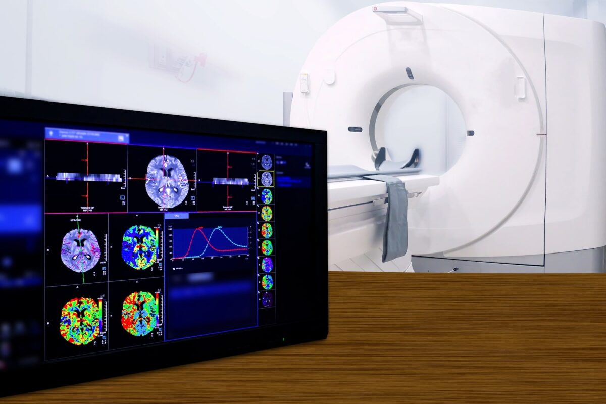
However, few patients who arrive late (beyond 4.5 to 6 hours and till 16-24 hours) can also be treated based on their imaging if we can identify salvageable penumbra (affected brain parenchyma but without permanent damage) using advanced imaging techniques that analyze brain parenchymal perfusion.

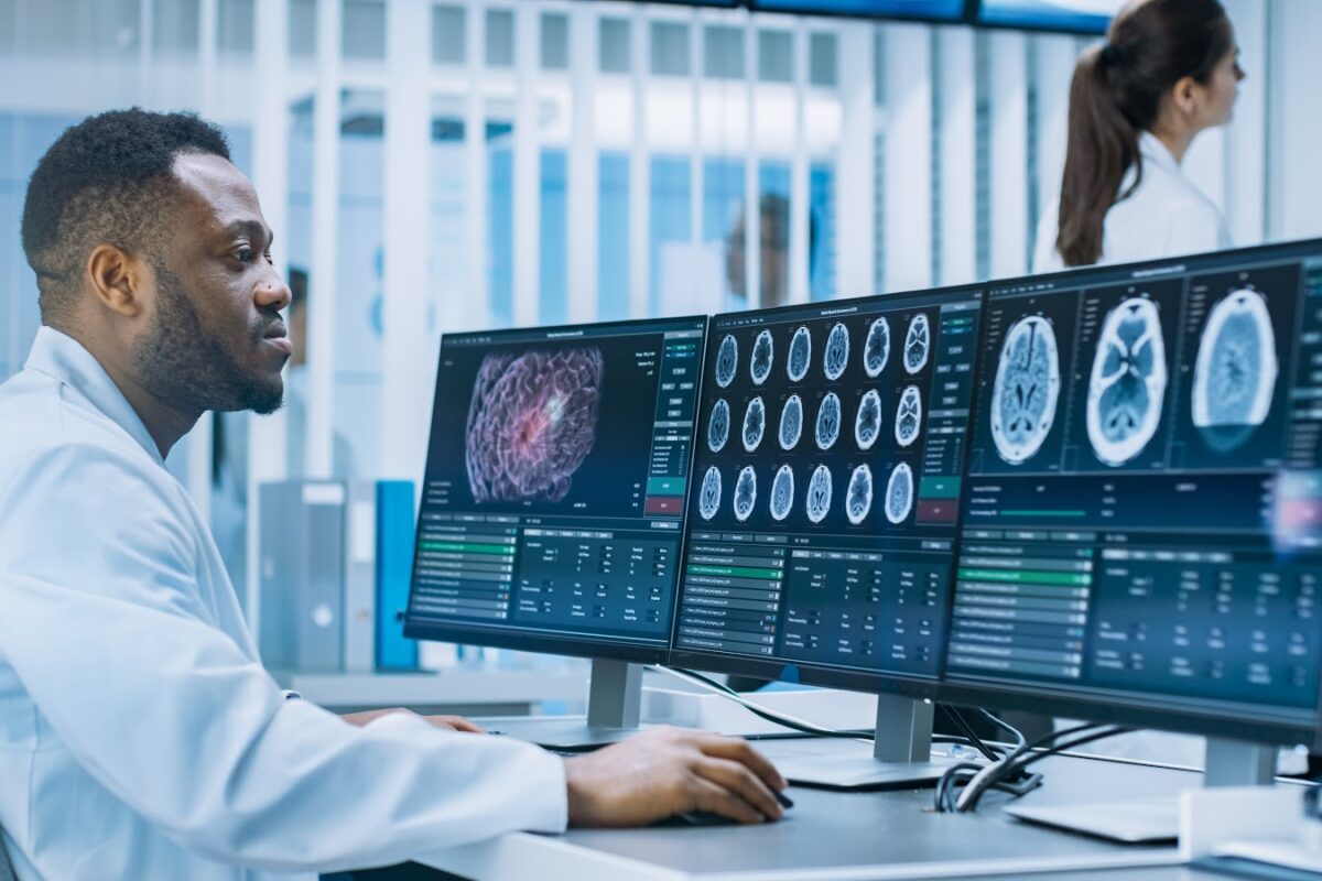
All of these decisions can also be taken collectively and more accurately using MRI. Ultrafast Stroke MRI takes less than 20 minutes using limited protocols and involves basic sequences as well as Angiography. MRI in acute stroke can be done without having to administer intravenous contrast and is especially very useful in differentiating from other mimics. Parenchymal and vascular information both can be obtained. MRI involves DWI for infarct core and MRA for LVO while FLAIR mismatch can be used for IVT in stroke patients with unknown time of onset or wake-up strokes. SWI can help detect thrombus/clot burden in RBC rich clots and serve as a penumbral marker by demonstrating vessels with raised oxygen extraction.

Teleradiology can thus be very effective in acute Stroke if reporting can be accelerated to avoid time delays. Simplified reporting formats can be developed based on local logistics. Although CT and CTA are widely practiced in acute stroke, MRI provides a non-contrast one-stop solution in the emergency especially if reported immediately by dedicated, trained tele-stroke Radiologists. Structured reports for such time-based emergency situations like acute stroke help avoid medico-legal consequences especially in the era of evidence-based medicine and helps guide the referring physicians in making treatment decisions.

References

1. Varadharajan S, et al. Computed tomography angiography in acute stroke (revisiting the 4Ps of imaging). Am J Emerg Med. 2016 Feb;34(2):282-7

2. Varadharajan S. ASAP In Stroke Imaging – Need to Simplify and Standardize in Emergency. Accepted for publication in Neurology India
Teleradiology Services India

Quality in Teleradiology Reporting

Quality is the extent to which the right procedure is done in the right way at the right time, and the correct interpretation is accurately and quickly communicated to the patient and referring physician.” Hillman et al

Teleradiology is playing a crucial role in delivering patient care with its prompt and quality service.

The quality of radiological reporting depends on multiple parameters apart from the internal review and external feedback system like

  • Communication between the reporting radiologists and referring physician for better understanding of the case
  • PACS and workflow management
  • Operational team (CTC)
  • Access to reporting tele-radiologist for clarification / discussion

The Internal Parameters can be listed as –

1. Internal Quality Assurance Team sets up an auditing system involving methodology on random review of the reports and peer reviews at regular intervals. The key performance indicators (KPIs) are prepared by the team in alignment with the organization’s objective and external auditor’s standards (if involved). The experts evaluate the reporting by carrying out random double reporting, peer review, and discuss the discrepancies with reporting radiologists.

Evaluation of the discrepancies, formulation, and implementation of corrective measures followed by evaluation of implementation is a cyclic process in order to improve the quality of report and service delivery. The option of contacting the reported tele-radiologist for further clarification and discussion on prognosis is available. 

The new recruits are initially engaged in preliminary reporting for familiarization with the tele-reporting system and are supported with constant review and feedback. This also involves eLearning, teleconferencing, telemedicine, and other technological processes.

2. Procedure Protocol indicates the Right Procedure & Right Way which impacts many other parameters. The protocol includes a checklist drafted by the team on imaging procedures, image uploading/transfer, availability of support documents, defined TAT for emergency & regular cases, filled request form, to & fro communication between the referring physician and reporting radiologists, assigning of images to radiologists, etc.

3. Credentials of the tele-radiologists including the sub-specialization and experience per se augment the value of the report. In critical cases like emergency / STAT reporting, the unique skill of picking up the hidden markers in a short time plays a critical or decisive role. Their clinical focus in reporting improves referring physician’s proficiency to advise or decide on suitable investigations/management of patients. 

4. TAT is variable and crucial for referring physicians in taking control of the situation, be it for emergency or regular cases. Today, because of the technological benefits, the TAT for an emergency case is approximately less than 30 minutes whereas for any inpatient or outpatient 4 to 8 hours is maintained. Especially in teleradiology, the other factor which determines the TAT is mutual agreement between the service provider & referring center and considering cost factor as well.

5.  Miss Rate is determined by continuous reviewing of reports at regular intervals by the review panel and a lot of measures are undertaken to keep it low. The positive culture of the review panel establishes an environment that is instructive, supporting cognitive debiasing and retrospective analyses. Clients are also encouraged to report “misses” by categorizing them based on their implicating severity. Feedback and discussions with referring physicians help in the process. 

The External Parameters which can be considered are

1.  Client’s feedback system serves two purposes – understanding the quality of report and reporting services although both (service provider and referring center) would have laid down the SOPs to be followed at either end. The upload of images of other recently conducted investigations, along with other laboratory, pathology reports and previous documents like discharge summary / clinical and operational notes helps in the accurate and detailed interpretation of the images.

2.  Accreditations by the authoritative body confirms compliance with protocols set by the authority matching the industry requirement and benefitting every stakeholder. Inspections are conducted to check the protocol implementation and service delivery at regular intervals and the certification is renewed accordingly. If any discrepancy is noticed, a Plan of Correction (POC) is provided with the timeline for implementation.

Cloudex Solutions ensuring the right procedure, the right way, right time, and correct interpretation

A team comprising of qualified experts like radiologists, clinicians, and technologists who are well supported with state-of-the-art RIS PACS technology, round the clock (24 x 7 x 365) reporting services complement to the client’s advantage.  The objective of doing the right thing at right time in the right way is achieved by integrating programs to adhere to protocol, utilize the quality improvement tools, and conduct of skill up-gradation workshops.  

Cloudex brimming with experts and a wide range of tele-reporting services ensures delivery of the correct interpretation at right time in the right way; enabling the healthcare service provider to deliver quality patient care without delay and assuring proper treatment saving on time and money.

At Cloudex, we support you to have that winning edge. 

Teleradiology in India

Teleradiology Reporting Services: Do You Really Need It? This Article Will Help You

The ever-evolving medical world keeps bringing out solutions to the challenges posed by the fast pace populace. Radiology is keeping pace with modernity and has very well integrated technology and telecommunication to formulate a remote mode of extending services – Teleradiology.

Today, Teleradiology has enabled the clinician in providing the best of patient care and saving lives and thus becoming an indispensable healthcare service. Teleradiology supports the clinicians/hospitals/healthcare service providers with many solutions, compensating the shortage of radiologists and reporting for all imaging modalities – X-Ray, Ultrasound scan, CT scan, MRI Scan, PET scan, Nuclear Medicine, Interventional imaging.

1. Round-the-clock Reporting service supports the healthcare service providers with radiology services round the clock for all days irrespective of no. of radiologists on-site or subspecialty reporting requirement.

The defined reporting TAT ensures a quick TAT of as early as 30 to 60 minutes for all modalities of imaging both in cases of emergency and regular cases. In this competitive and fast-paced environment, Teleradiology enables hospitals from remotest places to small or mid-size hospitals to provide patient care effectively irrespective of time.

2. Nighthawk Reporting services work out as an ideal option for many healthcare service providers who cannot afford to recruit a radiologist beyond regular working hours and get benefitted from after-hour radiology services.

Nighthawk reporting allows the radiology department to exhibit its duty of quality reporting without any delay. This late-night radiology coverage empowers the hospitals in extending patient care to a high level with better outcomes, and in a bigger perspective improves the face-value and bottom-line score of Hospitals. 

3. Subspecialty Reporting services help in identifying the subspecialty pathologies apart from identifying the common findings, normal variants quickly. The accuracy of reporting and detailed interpretation of the images (expertise in particular anatomy or type of reports/modality) improves the prognosis by facilitating the early diagnosis and proper treatment planning. Subspecialty reporting adds value eliminating the need for a second opinion; and improves the productivity and reporting TATs also.

Subspecialty reporting can be categorized into the following specialty

·       Thoracic Imaging

·       Breast Imaging

·       Cardiac Imaging

·       Abdominal Imaging

·       Musculoskeletal Imaging

·       Neuroradiology

·       Neuro-interventional Radiology

·       Vascular Interventional Radiology

·       Emergency Radiology

·       Pediatric Radiology

·       Nuclear Medicine

4. Emergency Reporting services ensures the early intervention and initiation of required action for emergency case. The involvement of expert radiologists reduces the TAT duration and contributes to the immediate diagnosis of acute conditions and patient care.

5. Weekend / Holiday Reporting services ensure the regular radiologists are able to utilize their break productively and return to job energized without any of the radiology reporting being stalked/trailed at the health center.

6. Second Opinion Reporting service is access to expertise for getting detailed imaging reports to facilitate a better understanding of the situation or decide on the course of treatment or to understand the progression of disease or treatment outcome. Experts are able to contribute through their experience and make a value-added difference to disease management/treatment.

7. Access to Radiology by healthcare centers present at geographically challenged areas either by altitude or distance. Primary or secondary healthcare centers have benefited from teleradiology which diminished the need for on-site radiologists and have benefitted from subspecialty reporting too. This has improved patient care services through quality and timing.

Benefits of Teleradiology reporting:

· Availability of radiology services round the clock (24 x 7 x 365) including night-time and on weekends/holidays

· Access to subspecialty radiologist’s reporting

· No backlog/delay in imaging reports delivery (improved TAT)

· High-quality patient care without delay

· Reports checked by more than one radiologist (preliminary/final reads)

· Better time management of radiologists due to login enabled cloud-based system (saves on commuting)

· Software enabled workflow helps in timely delivery of reports

Cloudex Radiology Solutions offers a plethora of Teleradiology Services:

The team of experienced, expert and qualified radiologists at Cloudex’s panel extend teleradiology services encompassing all type of reporting services to fulfill the growing demand of radiology services of health providers by defying the distances and time zone. The quick TAT, accuracy, standardized report protocol, coordination network between the reporting radiologists, clinician/surgeon/technician, and administrator contributes to the delivery of services benefitting the hospital and patient.

At Cloudex, we support you to have that winning edge. 

MUSCULOSKELETAL IMAGING

MUSCULOSKELETAL (MSK) IMAGING

Musculoskeletal Disorder (MSD), includes diseases of muscles, bones, joints, and the soft tissues around it. Pain and injuries are the most common complaints of the patients reaching out to a primary care physician, and with increasing age, the risk of developing MSD increases. As per the data from the World Health Organization (WHO), disability from MSDs is second largest in number, across the globe. The symptoms of various MSDs vary and it may affect the daily activity so much so that the quality of life may get compromised. It’s very important to get muscle or bone disorders diagnosed and treated in time, to keep oneself healthy. This makes MSK, the most sought after system as far as, an accurate clinical diagnosis is concerned.

Imaging Modalities

Various modalities are used to reach an optimum conclusion , before starting a confirmed treatment in MSDs. These are X-Rays, ultrasound, CT scans, and magnetic resonance imaging (MRIs). Each modality offers its benefits and limitations, that also, make them specific tools for diagnosis in specific health conditions. 

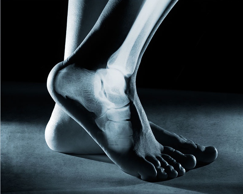
 X-rays are often performed in an emergency situation such as a traumatic fall or an accident leading to fractures. Limited information can also be received in conditions like arthritis, infections, and osteoporosis while using X-rays.

Ultrasound is used to take pictures of the anatomy and function of soft tissue around bones and joints using sound waves. Ultrasound imaging is typically used to diagnose tendon tears, muscle tears, fluid collections, ligament sprains or tears, inflammation or fluid within the bursae and joints, early changes of rheumatoid arthritis, nerve entrapments such as carpal tunnel syndrome.

CT scan is a procedure, which can produce detailed pictures of the muscles, bones, joints, and the soft tissues around. This painless and non-invasive procedure helps doctors making a diagnosis in cases of, Osteoporosis, Osteopenia, Paget’s disease, Bone cancer, Bone infections, Muscular dystrophy, Muscle sprains or strains, Tendinitis, etc.

● MRI (Magnetic Resonance Imaging)

MRI is the most sensitive non-invasive imaging tool for a broad range of diseases affecting the musculoskeletal system. MRI’s edge over the other imaging tools rests primarily in obtaining better image resolution of body tissues and pathologic processes. Thus, high-resolution images of MRI, bring the excellent and discrete view of organs and diseases.

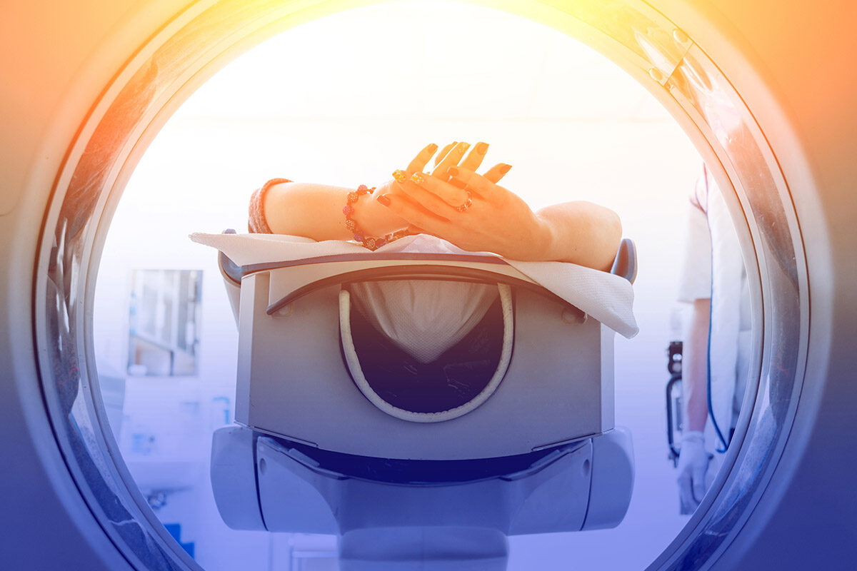
MRI uses a strong magnetic field, which is directly related to image resolution obtained. The field strength is typically between 1.5 T and 3 T, but may be lower in an open MRI scanner.

MRI is highly sensitive and specific for osseous and articular hip injuries, as well as soft tissues injuries of the hip and pelvis, and is the recommended imaging modality in most cases after a radiograph is negative.

Indications of MRI

Diagnostic imaging can be a valuable tool when used in conjunction with a thorough history and physical examination for establishing a diagnosis and treatment plan. Radiographs are the initial imaging modality in most cases of musculoskeletal injury or pain but MRI is most important and recommended imaging in following cases.

1.Bones

In the setting of trauma, MRI is indicated for the evaluation of those injuries of the metaphysis and epiphysis, where Xrays are found inconclusive/ undetected injuries, and to assess fracture union.

In knee injuries, When there is clinical suspicion for meniscal or ligamentous injury, MRI is the best modality to be used to detect such injuries.

MRI is useful to detect and stage acute and chronic osteomyelitis,primary bone tumors, both benign and cancerous, and undetected spread of secondaries to the bones.

2. Soft Tissues

 MRI is indicated for the evaluation of,

Muscle and tendon and ligament tears/injuries , hematomas, nerve entrapment syndromes,  tenosynovitis, bursitis, infections, abscesses and myositis  Rotator cuff injury (tendinopathy or tear), Avascular necrosis, Biceps tendon injury (tendinopathy/tear).

 It is helpful in detection, and staging of benign and malignant soft tissue tumours.

3. Joints/spinal

MRI is indicated for the diagnosis of hurt/ injuries to joints due to trauma, certain articular cartilage injuries, and inflammatory changes in the joints due to overuse, posttraumatic osteonecrosis and degenerative joint disease, joint infections, rheumatoid and the seronegative arthritis.

During traumatic spine surgeries, MRI is specifically recommended when there is a doubt for the injury to the spinal cord, nerve roots, and/or soft tissue, which are not well visualized on CT. MRI is also indicated in the cases of joint pain with clicking sounds, abnormal disc displacement, marrow abnormalities, osteonecrosis of the mandibular condyle and so on and so forth.

The list is endless……..and exhausting. Musculoskeletal health is very important to keep us going in our routine life, and in today’s day n time when the clinicians are overloaded and patients are becoming more impatient, there is a dire need for a diagnostic tool with an accurate, and in time reporting to have best results in day-to-day practice. MRI is the most recommended imaging tool in this scenario….

How do We Help….

At Cloudex, we have an entire team of certified Radiologists, Physicians, Subspeciality experts, and trained technologists, who work in association with Primary care  Physicians, Surgeons, orthopedic surgeons, and Radiation and Medical Oncologists to provide the best MRI reporting.

Our Fellowship Trained Experts from North America, provide structured reports based on the need and urgency. They are always available on call (24x7x365), for any discussion that can help managing any case, right from the simple fracture to an occult bone mass.

Urgent reporting within few hours, with upfront  information provided by the team to the referring doctor for various critical cases, has rewarded us with better patient and doctor satisfaction and immense faith of fellow clinicians.

Take Home Message from Cloudex……

We understand that the recipe to successful treatment results, is totally dependent upon the following ingredients:

The precision,

Reliability and,

In time access to an accurate, easy to understand imaging report,

At Cloudex, we ensure to help you have, that winning edge.

Teleradiology

BODY IMAGING

What is Body Imaging?

Body Imaging is a subspecialty of diagnostic radiology which deals with imaging evaluation of organs in the thorax, abdomen, and pelvis. It includes all types of radiological examinations performed using conventional X- rays, CT scan, MRI, ultrasound, mammography, and PET (positron emission tomography).