• +91 9544652090
  • support@cloudexrad.com
  • Working Hours - Mon - Sun: 12.00 am - 11.59 pm
What are you looking for?

Blog

4 STRATEGIES TO REDUCE REPORTING ERRORS IN RADIOLOGY

Reporting errors happen in radiology. Certainly they are not intentional, and many have little or no consequence to patients; others are significant. In an effort to be helpful, I would like to share four strategies to help reduce reporting errors in radiology. They are not intended to be in any way comprehensive, but they are actions that might help to increase accuracy.

It’s important to emphasize that I prefer the term “discrepancy” rather than error for much of what we’re considering. The term error implies a mistake, and that a clear-cut diagnosis and correct report are possible. However, as radiologists we know that there is not always a single definitive outcome from an imaging study. Imaging is rarely binary – “normal” or “abnormal”. We render an interpretation that is based on our understanding of the patient’s condition at the time of the exam. Often an “error” is determined later in the light of additional information and a developing clinical picture. The concept of necessary fallibility must be accepted. However, I will use the term “error” for the purpose of this blog, as it is the label most frequently used in literature and discussion.

How prevalent are errors? It seems that 3-5 % is the best minimum error rate achievable even when working in the best of circumstances (1). Knowing that one billion radiologic imaging exams are read annually worldwide, and assuming an average error rate of 4 percent, that equals approximately 40 million radiologist errors annually (2).

Strategy: address cognitive biases in radiology

We all have cognitive biases. They are the result of our brains’ attempts to simplify information processing. We cannot rid ourselves of these biases, but we need to be aware of them and take corrective actions to minimize their influence on our reporting (3). The following are only a selection of the many recognized biases to which radiologists are prone, with some suggested corrective measures of varying practical applicability. Admittedly, some of the suggested correction strategies are not feasible in usual radiology practice.

  • Anchoring bias – this is the tendency to rely on our initial impression and fail to adjust it in the light of subsequent information. Correction: avoid early guesses, and seek to disprove your initial diagnosis rather than confirm it. In some cases, you might want to get a second opinion.
  • Framing bias – this is the result of being influenced by the way a problem is framed. For example, if a referrer states, “the patient may have leprosy,” then your interpretation will be influenced by that statement, even though the likelihood of imaging findings being due to leprosy may be remote. Correction: initially review the study blindly before reading the clinical information.
  • Availability bias – this is the tendency to consider a diagnosis more likely if it readily comes to mind. For example, you are more likely to consider a pathology that you saw on a study the previous day, even if its likelihood is very small. Correction: try to use objective information to estimate the true base rate of that diagnosis, rather than relying on a quick initial impression.
  • Satisfaction of search – this is the tendency to stop searching for abnormalities once a likely diagnosis or first abnormality is found. Correction: use a systematic interpretation strategy, perhaps relying on a checklist or algorithmic approach, to help ensure a thorough review. Additionally, do a secondary search after initial abnormality detection, and also consider known combinations (e.g associated multiple injuries that commonly occur together in the knee).
  • Premature closure – this is the tendency to accept a diagnosis before full verification. Correction: always give a differential diagnosis. Never make a working diagnosis absolute without pathological confirmation. It’s important to make clear that I DON’T advocate this suggested corrective strategy; it would diminish the value created by radiology in patient care).

Strategy: probe for more patient information

I realize that it can be difficult to find time for clinical consultations with our referring colleagues, and for direct interaction with our patients. But I strongly believe that these activities are essential to improving our clinical practice. Also, their value is supported by several studies that show a higher percentage of errors occur when reporting is done by off-site reporters who had no opportunity to interact with the referrers or patients, and were presented with only a limited amount of clinical information (4). It is part of the job of radiologists to probe for more information when our instincts tell us the picture we have been given is incomplete.

A few helpful actions are:

• Discussing the appropriateness and justification of scans

• Tailoring studies to the specific clinical question

• Asking for appropriate missing snippets of history, rather than just proceeding because of time pressures

• Having direct discussions with referrers (including within multidisciplinary team meetings) about the significance of the scan results

Strategy: improve report writing

Sometimes we may interpret imaging studies accurately but be unclear in how we convey our meaning in the written report. From the patient’s perspective, the outcome can be the same whether we miss a potential diagnosis or we identify the relevant abnormalities but fail to effectively communicate the key findings and/or their meaning in a poorly-written report. If our reports are incoherent, rambling, and verbose – and if it’s impossible for the referrer to clearly understand what is most important in them – then we have failed to communicate, and are as guilty of “error” as if we missed the relevant findings entirely.

In fact, communication failure in general is the fourth most common reason for radiologists in the U.S. being sued, and 60% of these cases were due to a failure to highlight an urgent or unexpected abnormal finding and to emphasize it appropriately in reports.

I recommend you take a look at your own past reports with a fresh set of eyes, or perhaps ask a trusted colleague to read them. Look closely at your report structure, its organization, and your vocabulary choices. Are there mistakes in grammar or punctuation? Have you failed to correct errors in voice-recognition transcription, leading to confusion about your meaning?

I personally am not a huge fan of structured reporting, but I acknowledge that using them, especially for complex imaging studies, increases thoroughness and accuracy.

My recommendation: make your reports simple and clear, correct typographical errors, include what matters, do not include the irrelevant.

Strategy: ease mental and visual fatigue

Visual fatigue results from prolonged focusing on a workstation, and can be  alleviated (in part) by accommodative relaxation, shifting your visual focal point from near to far (e.g. looking at a distant object for 15-30 seconds) every 15 minutes.

Prolonged focus on a workstation causes visual and mental fatigue.

Mental fatigue is the consequence of continuous and prolonged decision making. We need to be aware that our cognitive processes respond to this mental strain by taking short cuts that might result in poor judgement and diagnostic errors. Here are a few suggestions that that might help you ease your mental fatigue (5):

  • Read the most difficult cases at the beginning of your shift when you are fresh.
  • Switch periodically between modalities.
  • Take structured breaks.
  • Reduce unnecessary interruptions and distractions

It is impossible to expect 100 percent accuracy 100 percent of the time, even under the best of circumstances. Our working environments in this current era of expected hyper-efficient radiology are far from ideal. Radiologist “error” may arise from personal issues, such as the visual and mental fatigue mentioned above, but systemic issues beyond our control (staff shortages, excessive workload, inadequate equipment, poor lighting conditions, lack of availability of previous studies etc.) are also frequent contributors, and they are unlikely to ever be completely eliminated.

Shifting to a system-centered view of errors

In addition to taking steps to minimize the occurrence of errors, we should also consider our reaction when they do happen. The traditional approach within medicine has been  person-centered, with errors viewed as indicative of a personal or professional failure. This culture of “naming, shaming and blaming” can result in suppression of error reporting as well as missed opportunities to learn from each other’s mistakes, and to make process improvements. We need understanding and support from each other and from others in healthcare when mistakes happen.

I believe we should shift our focus to the system, rather than the individual. A system-centered approach facilitates exploration of why an error happened and what can be done to prevent it from happening again. The National Radiology Quality Improvement Programme of the Faculty of Radiologists of the Royal College of Surgeons in Ireland is an example of an effort to embed in practice this more-enlighted and more-beneficial approach to errors. (6)

I also believe that we as a profession need to educate our patients about error rates. As leaders in radiology like Giles Maskell have emphasized, there is a yawning gap between what we know to be our error rate and what our patients believe it to be. The discovery in hindsight of an error in interpretation of a radiological image is often perceived by the patient as something shocking and exceptional, calling into question the competence of the radiologist and the overall care they are receiving. It would benefit radiologists if patients, referrers, and others in healthcare better understood the pervasive nature of radiological “error”, the inherent uncertainty in much of what we do,  and the measures we take to avoid it, while also emphasizing the enormous benefit that radiology – despite its inherent flaws – continues to bring to patient care.

In closing, I will share this quote from Sir William Osler, English/Canadian physician, who said, “Errors in judgement must occur in the practice of an art which consists largely in balancing probabilities.”

What are your thoughts and strategies for reducing errors in radiology? Please comment below.

This content was originally presented by Dr. Brady at ECR 2023.

Conclusion

Esr president Adrian Brady recently sat down with Carestream to discuss the 4 best  Strategies for reducing reporting errors in radiology.

We would like to thank https://www.myesr.org/ for providing valuable information and resources for this article.

DEVELOPMENT OF HEPATOCELLULAR CARCINOMA

VIRAL HEPATITIS

HBV and HCV are major causes of viral hepatitis that lead to the development of cirrhosis and HCC. HBV gains entry into liver cells through a receptor mediated pathway. HBV-DNA integration into host genetic machinery causing DNA methylation resulting in oxidative stress and formation of HBx protein(1). The risk of developing HCC has been shown to be proportional to HBV-DNA level in liver cells. Chronic illness results from persistence of the virus in the host cells via various mechanisms that include infection of immune defense control centers, viral inhibition of antigen presentation,selective immune suppression, down-regulation of viral gene expression, and viral mutations that functionally incapacitate virus-specific T cells from recognizing HBV antigen(2)

HCV hijacks host cellular machinery to increase cellular proliferation, steatosis, inflammatory processes, mitochondrial dysfunction, insulin resistance, all leading to oxidative stress, genetic instability and DNA damage with cirrhosis and HCC as a likely outcome(3)

NON-VIRAL HEPATITIS

Diabetes mellitus, alcohol abuse, cardiovascular disease, liver inflammation, obesity, dyslipidemia and non-alcoholic fatty liver disease (NAFLD) are some other major contributors to HCC development(4)

Accumulation of iron in the liver of NASH and HCC patientsis correlated with progression of fibrosis and HCC. NAFLD provides the metabolic environment to induce insulin resistance a known etiological factor for HCC(4,5).

Obesity impairs metabolism, induces inflammation and is an etiological factor for NAFLD, steatosis, NASH, hepatic fibrosis, cirrhosis, and ultimately HCC.

Toxic by-products of alcohol catabolism such as accumulation of acetaldehyde and free radicals can influence oxidative stress, apoptotic cell death, necrosis and necroptosis(6).Reactive oxygen species (ROS) generation is the result of increased inflammatory cytokine. ROS-induced DNA damage, genomic vulnerability of hepatocytes and T-lymphocyte suppression contribute to HCC development. Alcohol diet have shown exacerbation of inflammation, epithelial-mesenchymal transition (EMT) andfibrosis, and consequent progression to HCC (3).

Other possible risk factors include genetic predisposition and congenital abnormalities, toxic exposures (aflatoxin or arsenic contaminated food), and autoimmune diseases of the liver. The pathogenesis of aflatoxin B1 (AFB1) – induced HCC includes several mechanisms, including the formation of mutagenic and carcinogenic intermediates and adducts. These adducts and intermediates can also directly induce a mutation at codon 249 of the p53 tumor suppressor gene. This replaces arginine with serine, a change that reverses the tumor suppressing ability of the gene. There are reports that suggest that AFB1 acts synergistically with HBV to induce HCC(7).

DIAGNOSIS OF HEPATOCELLULAR CARCINOMA

IMAGING MODALITIES

ULTRASOUND:

  • Small focal HCC appears hypoechoic compared with normal liver.
  • Larger lesions appear heterogeneous due to fibrosis, fatty change, necrosis and calcification.
  • A peripheral hypoechoic halo may be seen with focal fatty sparing
  • Contrast-enhanced ultrasound 1

o arterial phase-arterial enhancement from neovascularity

o portal venous phase

 Decreased echogenicity relative to background liver ie wash out.

 Tumour thrombus may be visible.(8)

Figure 1:Two different HCC lesions (arrows) in gray-scale ultrasound (a,c)
and in late phase of contrast-enhanced ultrasound (b,d)(9)

CONTRAST ENHANCED MULTIPHASE CT:

Imaging protocols are

  1. The patient was positioned in the supine position.
  • Technical parameters were X-ray tube current 160 to 220 mA; tube voltage 120 kV;

collimation 5mm; rotation speed 0.75 s; matrix 512×512. Iohexol (350 g/L) was used to

perform the contrast-enhanced scanning.

  • A high-pressure syringe was used to inject 1.2-2 ml/kg of contrast agent at injection rate

of 3.5- 4.0mL/s. Twenty millilitre saline were later injected at the same rate.

  • Scanning range was set to from the lower chest to the to lower abdomen level.
  • All image data were transmitted directly to our picture archiving and communication

system. Sagittal and coronal reformats of images were also obtained(10).

Imaging of FLLs in CT requires the use of a multi-phase study protocol.

  • Includes a phase prior to the intravascular administration of contrast agent.
  • Phases obtained after intravascular administration of contrast medium – Hepatic arterial

phase(HAP), Portal Venous Phase(PVP) and Equilibrium phases(EP) obtained routinely

40, 60 and 180 seconds post contrast administration respectively in a multi-row CT unit

  • EP may be also referred to as an early delay phase in comparison to the late delay phase,

obtained after 10 to 15 minutes after administration of contrast medium, acquired if the

imaging protocol is extended to detect lesions with a high content of fibrous tissue(9).

Figure 2:Multiphase CT. Native examination (a), hepatic arterial phase with contrast agent in
hepatic arteries and slight enhancement in portal vein (b), portal venous phase (c) equilibrium
phase (d).

Imaging features in Hepatocellular carcinoma

  • HCCs enhance strongly in the HAP, depending on the size of the tumor and the presence

of regressive changes homo- or heterogeneously. Large tumors will typically present with

heterogeneous enhancement, often with so-called mosaic pattern as opposed to small,

early forms of hepatocelullar carcinomas

2. Washing-out of the contrast agent in PVP (the phase of the strongest enhancement of the

liver parenchyma) or/and EP is a sine qua non for diagnosing HCC with specificity of 95-

96%.

3. If tumor pseudo-capsule is present, it is more clearly visible in the PVP and EP than in

HAP, with delayed enhancement in EP. Tumors with pseudo-capsule show better

prognosis(11)

Figure 3: Schematic presentation of pattern of enhancement of HCC lesion with strong enhancement
in HAP and wash-out of contrast agent in subsequent PVP and EP(12)
Figure 4:CT axial images. HCC in hepatic arterial phase (a) and equilibrium phase (b). Wash-out
feature and enhancing tumor pseudo-capsule is visible in the latter(9).

DYNAMIC CONTRAST ENHANCED MAGNETIC RESONANCE IMAGING

Standard MRI protocol consists of

  • Patients were positioned supine head first on the MRI table, then the MRI was

performed including T2 weighted fast spin-echo (T2-FSE) and DIXON, Duel echo

sequence(TE 90 and 180 ms), chemical shift imaging (in- and opposed-phase) and

diffusion-weighted image (DWI) map was performed for using b value of 500 (with

TR 1300 ms & TE 64 ms), with corresponding ADC mapping.

  • CE-MRI was performed afterward using gadopentetate dimeglumine

(Omnivist)/gadoterate meglumine(Clariscan) injected through an antecubital

intravenous catheter at a rate of 1.2 ml/min over 15 s and a dose of 0.2 ml/kg followed

by saline chaser of 20 ml at a rate of 1–2 mL/s

  • Dynamic contrast-enhanced sequences were acquired using DIXON sequence

acquired before (pre-contrast) and after contrast injection at 15-20 s (arterial phase), 40

s (portal phase), 60s (venous phase), and 180s (delayed phase). All contrast sequences

were acquired at the axial plane(13).

Imaging features in Hepatocellular carcinoma

  • Pre-contrast MRI sequences the majority of large HCCs show decreased signal intensity

in T1-weighted and increased signal intensity in T2-weighted images.

Small lesions tend to remain isointense to the adjacent liver parenchyma in T1-weighted

images Presence of intracellular fatty components may be easily confirmed in phase and

out of phase sequences.

  • Decrease in signal intensity in T2-weighted images is seen in case of fibrous tissue,

while areas of necrosis present especially within large foci cause an increase in signal

intensity and lead to heterogenous enhancement.

  • Low signal intensity of regenerative nodules in T2-weighted images resulting from

characteristic iron deposits, facilitate differential diagnosis with usually hyperintense

HCC foci.

  • Pseudocapsule is hypointense in T2-weighted images and shows delayed enhancement

in EP, similarly to CT.

  • DCE-MRI shows a similar enhancement pattern in majority of HCCs as observed in

multiphase CT with early strong enhancement in HAP and washing-out in the following

phases.

Figure 8:

Stepwise carcinogenesis of HCC in cirrhosis

Table 1: Various imaging appearance of cirrhotic nodules to frank HCC in MRI
  • Early washout can be seen in high grade or undifferentiated hepatocellular carcinoma

since the lesion is entirely supplied by hepatic artery. So the phase of washout will

help in diffentiation the grade of hepatocellular carcinoma.

  • Hepatocellular carcinomas have fat within it where as dysplastic and regenerative

nodules do not contain fat within it.

  • HCC directly invades the vessel and enhances on arterial phase.
  • Collateral formation and prominent adjacent vessel are the additional imaging features seen in HCC.
  • Intratumoural psuedoaneurysms are common in HCC(9).
Figure 5:Large HCC with degenerative changes in coronal T1-weighted image with fat saturation

(a) and in coronal T2-weighted image (b). Dynamic contrast-enhanced sequences in axial T1-

weighted images with fat saturation after administration of hepatocyte-specific contrast agent in

hepatic arterial phase (c), portal venous phase (d) and hepatobiliary phase (e). Heterogenous

enhancement of the lesion is seen with areas of non-enhancing focal necrosis (c) with subsequent

washing out of the contrast agent (d). Lesion shows low signal intensity in comparison to adjacent

liver parenchyma in hepatobiliary phase (e) (9).

OTHER PARAMETERS FOR DIAGNOSIS OF HCC

LABORTARY TESTS-Serum AFP is the most widely used tumor biomarker in

diagnosis of HCC. An increase of serum AFP levels in cirrhotic patients, however its

value is often considered insufficient(14)

HISTOPATHOLOGY- Well vascularized tumors with wide trabeculae (> 3 cells),

prominent acinar pattern, small cell changes, cytologic atypia, mitotic activity, vascular

invasion, absence of Kupffer cells and the loss of the reticulin network(12).

ctchest teleradiology

Teleradiology Services Are Forerunners Among the Healthcare Providers to Adapt to Hybrid Work in Contemporary Times

The healthcare management faced a tough situation during a pandemic time and emerged victorious in extending the best care to COVID & Non-COVD patients as well as safeguarding its medical care providers (clinical & non-clinical); and with the understanding that technology-driven practices and contact-less patient care are the new norms and necessity. 

Today, medical technology integrated with deep learning and artificial intelligence is one of the major contributing factors. On-line consultation through telemedicine, teleradiology, drone services, e-pharmacy, remote working, and at home services are some of the examples contributing to reduced or no exposure. Radiology was among the forerunners that made a swift adaption to new norms and supported the system with remote working. The technology companies too supported the medical system’s functionality either in maintaining or increasing the efficiency of the operational team during the patient flow surge. 

Teleradiology system driven by technological advancement has emerged as one of the most reliable modes of the patient care system in any given situation to deal with, be it a reporting for emergency or isolated COVID patients or IP/OPD patients, and could cope with the surge in image reporting through the option of remote reporting. This hybrid work norm supported many institutions in delivering patient care without any delay. Today’s teleradiology is integrated with the latest PACS, AI enabled workflow, internet connectivity with wide bandwidth and an experienced back-end support team that has made the reporting possible with short TAT. Technology companies have developed systems to transfer and archive the large dataset with integrity fulfilling the radiologist’s need for high-quality images for reporting. 

Machine Manufacturers are also contributing by developing systems that capture high-quality images with low dose radiation and high sensitivity. And to catch up with the trend of imaging services at point-of-care, compact mobile machines are getting developed to fulfill the need. The ultrasound machine and mobile X-ray machines are proving it with their wire-less and cloud-computing features. 

Remote Learning is being embraced by educators and many clinicians. The online recorded or live sessions have become the mode of learning for aspiring future physicians and medical professionals. Webinars/conferences and interactive sessions are being conducted virtually benefiting the healthcare providers and patients. 

With progressing time, the positive and productive approaches of radiologists, healthcare providers and technology companies the recovery from the pandemic is becoming a reality.  

Cloudex Teleradiology Solutions during Pandemic

Cloudex with its advanced teleradiology service in India supported many health centers worldwide managing the patient surge with its prompt reporting services. The reporting was dominated by X-rays, CT and MRI scan mainly for emergency cases as other patients were curtailed from outpatient services. Our experienced and expert teleradiologists team extended their much needed services relentlessly round the clock. Safety measure guidelines were strictly followed by the back-end team while reporting was done remotely. The non-stop and timely service deliverance which includes subspeciality reporting in this crucial time helped many centers in facilitating patient recovery and saving lives and it turned out as their winning edge. 

At Cloudex, we support you to have that winning edge.

teleradiology

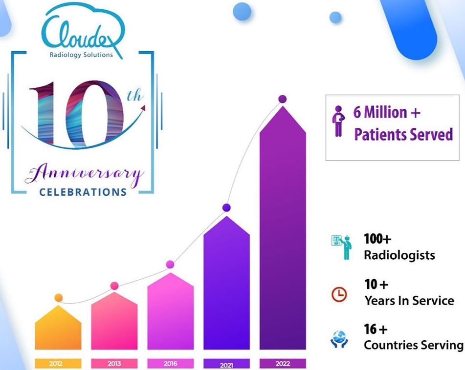
Cloudex Celebrates its 10 years of Teleradiology with 6 million patient’s smile curves

A humble start of Cloudex’s teleradiology services in 2012 has grown in leaps and bounds and has reached this celebration point. The journey so far covered has been full of coordination, compassion and patience filled. Each day has passed by with learning, growth and sometimes stumbling’s too. Today, we can share and write on our success stories; and redefine it with growing service demand, advancing technology and addition of sharper minds.

Our success is because of our dedicated team who nurtured Cloudex’s initiation into international presence, helped in soaring the service avenues and winning many hearts and clients.   

The intention of contributing to community has been formulated with care in order to bridge the gap in radiology services – learning & skill upgradation programs apart from setting up radiology service centres and teleradiology service support to healthcare centres. Cloudex has initiated online and offline developmental programs for aspiring radiology clinicians, expanding its service network through partnership beyond boundary. We are intensifying our venture in improving accessible radiology by establishing scan centres under PP partnership where increasing patient scan numbers talks about our investment on people, technology, equipment and customer service.

Our Milestone achievements in past 10 years can listed as

  • Over 6 million patients benefitted with our services
  • Over 100 Radiologists contributing to the smile curve
  • International presence of 16+ countries
  • Team of 40+ roped together with one mission of service

Currently, Cloudex is extending its clinical service to healthcare centres across India including those located in difficult to reach terrains and rural parts. And internationally, some of the centre in African countries like Botswana, Ghana, Ethiopia, Sudan, Nigeria, Uganda, Kenya, Tanzania and Southeast Asian countries like Malaysia, Philippines, Indonesia, Thailand, Vietnam are benefitting from our services.

In this demanding pandemic time, empowering the health centres with the winning edge seems to be success mantra. And Cloudex accepts it and strives to remain at par with industry trend, match the expectations of the associated healthcare service providers, raise the service delivery standards. And create a conducive work environment for the team which propels to give their best efficiently. At Cloudex, we strive to provide that winning edge or satisfaction and saving life.

ct scan_workshop

5 Influencing Factors in Teleradiology Reporting

Radiology reporting with shorter TAT aided with tele reporting has brought about significant improvement in patient care outcomes in many departments of medical centers. TeleRadiology report plays a crucial role in terms of diagnosis and treatment planning. And sometimes, a few minutes of waiting period feels like long years and one wonders how to overcome those anxious moments of dilemma. Silence prevails on the patient’s side whereas an array of actions happens on the other side for radiology reporting. So, behind the complexity of radiological image interpretation lies an array of actions involving technology, protocols, coordination, communication, expertise which keep happening seamlessly. Many of these are visible in action status at the reporting stations or work stations of radiologists (on-site/off-site).

The radiologist/teleradiologist’s work-station is made up of hardware, software, and appropriate furniture & interiors. Factors like illumination in working space, ergonomics of chair & table to smallest of an object like a mouse which can possibly trigger the discomfort level to radiologists matters in the process. Briefing out these dynamic factors for understanding behind the scene actions in teleradiology –

1. Hardware:

  • The dual QHD monitors of size 21 / 30” diagonally equivalent to a 3 or 5 megapixel are used to view the images and write reports simultaneously. This monitor size reduces the need for repeated zooming in or panning out.
  • CPU with 8th generation, 16 GB RAM, 1GB GPU memory to match the speed of the image interpretation process of the radiologist.
  • V shaped keyboard and optic mouse with a scroll which contours the fit into adult hand providing the ergonomic posture for convenient working.
  • Bluetooth enabled headsets helps in audio and dictation.

2. Software:

  • PACS provides seamless transmission of images in correct order along with other required supporting health documents.
  • DICOM calibration to meet the DICOM standards which ensures the image displayed is perceptually linearized. This software also provides alerts for out-of-compliance issues through emails/notification.
  • Notification system alerting the radiologists on an upcoming critical case that needs urgent attention, TAT alarm, call out from other physicians for case discussion/clarification.

Communication platforms contribute in an immeasurable manner. The interaction between the reporting teleradiologist and referring physician is crucial and is a deciding factor and lifesaving in many cases. The alerts/notification / textual or audio messages etc have added speed to the reporting system.

  • TAT enhancing voice recognition services add lot of value to teleradiology services. It enables teleradiologist in engaging more time in image interpretation and sparing them from initial drafting of reports with features like Real-time self-editing, formatting, auto-punctuation, and electronic signature. It works as workflow accelerator helping in completing the greater number of image reporting in given time frame.

3. Furniture & Interiors:

  • The versatility of working space supports the radiologist’s long working hours. The ergonomics of the furniture like height of a table, lumbar supporting chair with adjustable seat height are important for comfortable viewing and reporting. This subtlety adds in promoting alertness and efficiency.
  • Luminance is given special care so as to safeguard radiologists from ocular distress and physical strain. The minimum standards of 350 ccd/m2 is maintained.

4. Connectivity & Server: Speed is the vital part because a lot of activity depends upon it – transmission, downloading, and storage-related rendering or archival of images, interpretation, and reporting of images.

  • A dedicated reliable high-speed internet connectivity is maintained. The minimum required internet speed is 512 KBPS-1MBPS whereas the speed of 4-8 MBPS is considered ideal enabling a smooth process for interpretation & reporting, and desirable speed is above 25 MBPS for interpretation of large image data set.
  • The whole process is supported through cloud computing or a quality-controlled web server

5. Cybersecurity: The infrastructure of PACS & DICOM are build keeping in mind the various guidelines provided by HIPPA/FDAs which are further integrated or upgraded with mitigation measures decided by the Teleradiology companies as company policy or SOP decided upon with their clients. Some of the well-known measures are selective encryption of the DICOM header/files, transport security, image de-identification, water-marking or digital signatures, and access to the system thru assigned user ID/password, etc.

Cloudex’s user-friendly work stations delivering customer satisfying results

At Cloudex, reporting stations are configured involving lots of detailing to ensure that teleradiologists are able to concentrate on interpretation and reporting without any distractions/interruptions and operational issues. The reporting platforms are integrated with various software like voice-recognition dictation, multimedia communication, notification apart from the latest PACS & case managing RIS. Importance is also given to illuminance, work-space comfort, sitting ergonomics, monitor configuration for maintaining teleradiologit’s optimal concentration and efficiency. We invest beyond infrastructure too for creating a conducive environment that is free from administrative burdens or logistical hurdles for teleradiologists. The organization’s emphasis on accuracy of reporting without a miss at required TAT facilitates in winning the confidence of referring Physician and scoring high on service satisfaction score.

Cloudex with its wide range of teleradiology services – Emergency, Subspecialty, Nighthawk, Round-the-clock, Weekend / Holiday, Second Opinion; enables the referring physician or medical center in having an edge of being quick, effective, and efficient in delivering patient care. At Cloudex, we support you to have that winning edge.